| 中文名称 | 磷酸化肌球蛋白调节多肽9(平滑肌亚型)抗体 |
| 别 名 | MYL9 (phospho S20); p-MLC(Ser20); phospho-MLC(Ser20); p-Myosin light chain(Ser20); MYL9_HUMAN; Myosin regulatory light polypeptide 9; 20 kDa myosin light chain; LC20; MLC-2C; Myosin RLC; Myosin regulatory light chain 2, smooth muscle isoform; Myosin regulatory light chain 9; Myosin regulatory light chain MRLC1; MLC2; MRLC1; MYRL2. |
| 产品类型 | 磷酸化抗体 |
| 研究领域 | 心血管 信号转导 干细胞 细胞骨架 细胞膜蛋白 |
| 抗体来源 | Rabbit |
| 克隆类型 | Polyclonal |
| 交叉反应 | Human, (predicted: Mouse, Rat, Dog, Pig, Cow, Rabbit, Sheep, ) |
| 产品应用 | WB=1:500-2000 ELISA=1:500-1000 IHC-P=1:100-500 IHC-F=1:100-500 IF=1:100-500 (石蜡切片需做抗原修复) not yet tested in other applications. optimal dilutions/concentrations should be determined by the end user. |
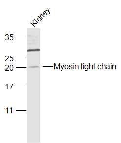
| 分 子 量 | 20kDa |
| 细胞定位 | 细胞浆 |
| 性 状 | Liquid |
| 浓 度 | 1mg/ml |
| 免 疫 原 | KLH conjugated synthesised phosphopeptide derived from human MYL9 around the phosphorylation site of Ser20:AT(p-S)NV |
| 亚 型 | IgG |
| 纯化方法 | affinity purified by Protein A |
| 储 存 液 | 0.01M TBS(pH7.4) with 1% BSA, 0.03% Proclin300 and 50% Glycerol. |
| 保存条件 | Shipped at 4℃. Store at -20 °C for one year. Avoid repeated freeze/thaw cycles. |
| PubMed | PubMed |
| 产品介绍 | Myosin light chain (MLC) is a subunit of the conventional myosins (e.g. myosin II). In smooth muscle and non-muscle cells conventional myosins mediate a wide variety of contractile events including cytokinesis, cell motility, and smooth muscle contraction. MLC is phosphorylated by multiple serine-threonine kinases such as Rho-kinase and PAK, however myosin light chain kinase (MLCK) acts as the primary kinase. Contractile activity of conventional myosins is regulated by phosphorylation of MLC on several residues. Subunit: Myosin is an hexamer of 2 heavy chains and 4 light chains. Tissue Specificity: Smooth muscle tissues and in some, but not all, nonmuscle cells. Similarity: Contains 3 EF-hand domains. SWISS: P24844 Gene ID: 10398 Database links:
Entrez Gene: 10398 Human Entrez Gene: 98932 Mouse Entrez Gene: 296313 Rat Omim: 609905 Human SwissProt: P20689 Human SwissProt: P24844 Human SwissProt: Q9CQ19 Mouse SwissProt: Q64122 Rat Unigene: 504687 Human Unigene: 271770 Mouse Unigene: 228729 Rat Unigene: 6870 Rat
Important Note: This product as supplied is intended for research use only, not for use in human, therapeutic or diagnostic applications. |
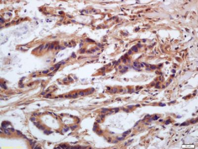
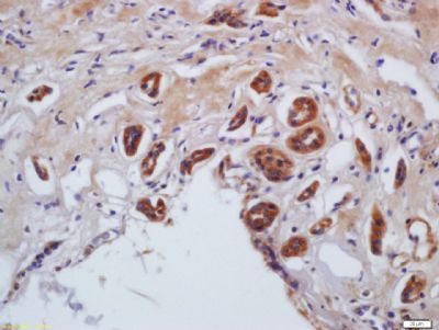
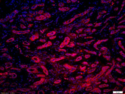
| 产品图片 |  Sample: Sample:Kidney (Mouse) Lysate at 40 ug Primary: Anti-Myosin light chain (phospho S20) (bs-7052R) at 1/1000 dilution Secondary: IRDye800CW Goat Anti-Rabbit IgG at 1/20000 dilution Predicted band size: 20 kD Observed band size: 20 kD  Tissue/cell: human gastric carcinoma; 4% Paraformaldehyde-fixed and paraffin-embedded; Tissue/cell: human gastric carcinoma; 4% Paraformaldehyde-fixed and paraffin-embedded;Antigen retrieval: citrate buffer ( 0.01M, pH 6.0 ), Boiling bathing for 15min; Block endogenous peroxidase by 3% Hydrogen peroxide for 30min; Blocking buffer (normal goat serum,C-0005) at 37℃ for 20 min; Incubation: Anti-phospho-MLC(Ser20) Polyclonal Antibody, Unconjugated(bs-7052R) 1:200, overnight at 4°C, followed by conjugation to the secondary antibody(SP-0023) and DAB(C-0010) staining  Tissue/cell: human kidney tissue; 4% Paraformaldehyde-fixed and paraffin-embedded; Tissue/cell: human kidney tissue; 4% Paraformaldehyde-fixed and paraffin-embedded;Antigen retrieval: citrate buffer ( 0.01M, pH 6.0 ), Boiling bathing for 15min; Block endogenous peroxidase by 3% Hydrogen peroxide for 30min; Blocking buffer (normal goat serum,C-0005) at 37℃ for 20 min; Incubation: Anti-phospho-MLC(Ser20) Polyclonal Antibody, Unconjugated(bs-7052R) 1:200, overnight at 4°C, followed by conjugation to the secondary antibody(SP-0023) and DAB(C-0010) staining  Tissue/cell: human kidney tissue;4% Paraformaldehyde-fixed and paraffin-embedded; Tissue/cell: human kidney tissue;4% Paraformaldehyde-fixed and paraffin-embedded;Antigen retrieval: citrate buffer ( 0.01M, pH 6.0 ), Boiling bathing for 15min; Blocking buffer (normal goat serum,C-0005) at 37℃ for 20 min; Incubation: Anti-phospho-MLC(Ser20) Polyclonal Antibody, Unconjugated(bs-7052R) 1:200, overnight at 4°C; The secondary antibody was Goat Anti-Rabbit IgG, Cy3 conjugated (bs-0295G-Cy3)used at 1:200 dilution for 40 minutes at 37°C. DAPI(5ug/ml,blue,C-0033) was used to stain the cell nuclei |
